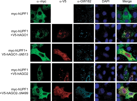
FIG. 5.
Subcellular localization of hUPF1. HeLa cells were transiently transfected with plasmids expressing hUPF1 and/or Argonaute proteins and stained with anti-myc (9E10), anti-V5, and anti-GW182 antibodies. The transfected plasmids are indicated on the left. Alexa Fluor 488-goat anti-mouse IgG, Alexa Fluor 594-goat anti-rabbit IgG, and Alexa Fluor 647-goat anti-human IgG were used as the secondary antibodies.

