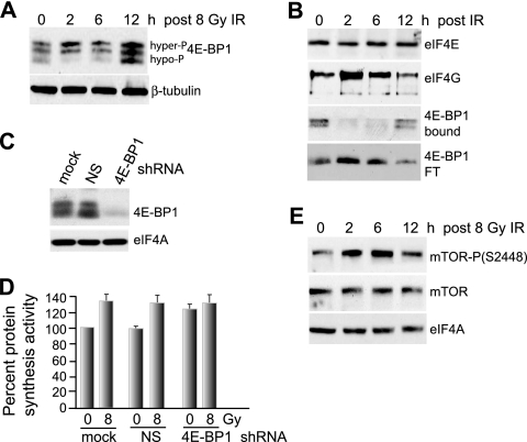
FIG. 3.
Increased hyperphosphorylation of 4E-BP1 early after IR promotes eIF4F complex formation and protein synthesis. (A) 4E-BP1 was resolved by high-resolution SDS-15% PAGE using equal amounts of protein extracts obtained from MCF10A cells irradiated with a single fraction of 8 Gy IR and harvested at the indicated time points. Hyper- and hypophosphorylated forms of 4E-BP1 are indicated. (B) Cells irradiated at 8 Gy were harvested at the times shown, equal amounts of protein extracts were subjected to m7GTP-Sepharose cap chromatography, and retentates were recovered by elution with SDS, resolved by SDS-PAGE, and detected by immunoblot analysis with antibodies as indicated. The flowthrough fraction (FT) of 4E-BP1 that did not bind to m7GTP-Sepharose is shown. The data were quantified by densitometry of autoradiograms from at least three independent experiments. Representative results are shown. (C) MCF10A cells were stably transformed with lentivirus vectors expressing NS or 4E-BP1-silencing shRNAs, and equal amounts of protein lysates were subjected to immunoblot analysis with antibodies as shown. (D) MCF10A cells silenced as for panel C were subjected to 8 Gy irradiation, and protein synthesis rates were determined 2 h later by metabolic labeling with [35S]methionine/cysteine. (E) mTOR abundance and activating phosphorylation at Ser2448 were determined in MCF10A cells irradiated at 8 Gy at the times shown by immunoblot analysis using specific antibodies as shown. All results are representative of at least three independent experiments, with standard errors of the mean determined from all studies.

