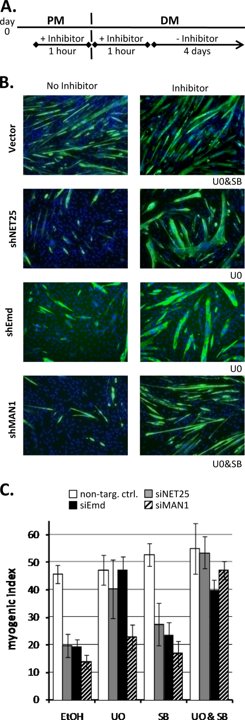
FIG. 7.
Pharmacological rescue of myogenic defect in C2C12 cultures after depletion of NET25, emerin, and MAN1. (A) Schematic of the time course of inhibitor application. On day 0, proliferating C2C12 cultures were treated with an inhibitor for the period between 1 h before and 1 h after shifting cells from PM to DM. Cultures were then kept for 4 days in DM before MI analysis. (B) Immunofluorescent micrographs (green, MyHC; blue, DNA) of empty vector control (Vector) and NET25 (shNET25)-, emerin (shEmd)-, or MAN1 (shMAN1)-depleted C2C12 cultures treated with MEK1/2 inhibitor U0126 (U0) and/or TGF-β inhibitor SB431542 (SB). The inhibitor applied to the drug-treated cultures (right column) is indicated below the bottom right corner of each image. Cultures were assayed after 4 days of differentiation. Images shown are from one representative experiment. (C) MI analysis of NET25 (gray bars)-, emerin (black bars)-, and MAN1 (hatched bars)-depleted as well as control (open bars) cultures treated with U0 and/or SB. Average values from the results for at least three independent experiments are shown. Error bars indicate standard deviations.

