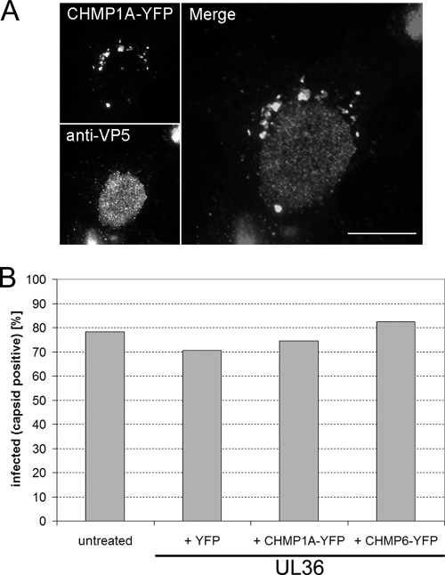
FIG. 4.
Overexpression of dominant-negative ESCRT-III proteins does not affect entry of HSV-1. COS-7 cells were either left untreated or cotransfected with a plasmid expressing UL36 together with a plasmid expressing CHMP1A-YFP, CHMP6-YFP, or YFP. Cells were infected at 24 h posttransfection with HSV-1 KΔUL36 at 5 PFU/cell. Cells were fixed 15 h postinfection, permeabilized and incubated with a monoclonal antibody specific for the HSV-1 major capsid protein (VP5), followed by an Alexa Fluor 568-conjugated anti-mouse secondary antibody. (A) Epifluorescence image of an infected CHMP1A-YFP-expressing cell. CHMP1A-YFP-positive cytoplasmic compartments can be observed (top left-hand panel) in a cell with high levels of VP5 in the nucleus (bottom left-hand panel), indicating that HSV-1 can enter cells and initiate viral gene expression in the presence of a dominant-negative CHMP. Right-hand panel shows merged image. Scale bar, 20 μm. (B) Cells were counted and scored for the simultaneous expression of YFP-tagged proteins and a characteristic nuclear staining of the HSV-1 major capsid protein (VP5), as an indication of virus entry and subsequent viral gene expression. Bars indicate the percentage of YFP or CHMP-YFP-expressing cells that were also VP5 positive from a total of 200 cells or the percentage of all cells that were VP5 positive from a total of 400 cells in the untransfected sample.

