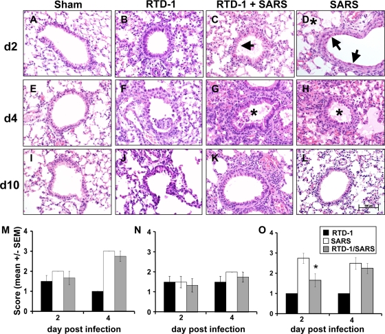
FIG. 2.
(A to L) Pulmonary tissue histopathology in SARS-CoV (3 × 10⁵ PFU MA15)-infected mice with or without RTD-1 (5 mg/kg, two doses) treatment. Hematoxylin- and eosin-stained (4 μM) tissue sections were examined 2, 4, and 10 days postinfection. See the text for additional details. Asterisks indicate alveolar edema; arrows indicate necrotizing bronchiolitis. n = 4 at each time point. Scale bar, 100 μm. (M to O) Pulmonary histopathology scores in SARS-CoV-infected mice treated with or without RTD-1. Tissues were harvested at 2 and 4 days and scored by a veterinary pathologist (D.K.M.) blinded to the treatment protocol, using a severity scale from 1 (absent/rare) to 3 (severe/multifocal). Data are presented for alveolar edema (M), perivascular cellular infiltrates (N), and necrotizing bronchiolitis (O). Results are presented as means ± standard errors (n = 4; *, P ≤ 0.05).

