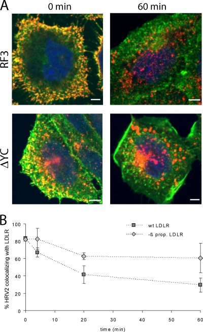
FIG. 3.
HRV2 dissociation from LDLR is delayed when the β-propeller is deleted. HRV2 was bound at 4°C to CHO cells grown on coverslips, and entry was initiated by adding warm medium. At the times indicated, the cells were fixed and permeabilized, and LDLR and HRV2 were detected by specific antibodies, followed by Alexa 488-conjugated goat anti-mouse IgG and Alexa 568-conjugated goat anti-chicken IgG, respectively. (A) Representative fluorescent images of one focal plane through the perinuclear region are shown after HRV2 binding (0 min) and 60 min after warming to 34°C. LDLR, green; HRV2, red. (B) The percent colocalization of virus and receptor was calculated from immunofluorescence microscopy images as in panel A. Colocalization at time zero was set to 100%. Error bars indicate the means ± the SD (n = 3).

