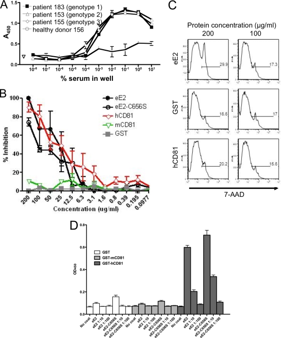
FIG. 7.
Functional analysis of eE2 and eE2-C656S. (A) ELISA plates were coated with eE2 and probed with a series of 10-fold dilutions of serum from patients infected with HCV (genotype 1, 2, or 3) or a healthy donor. Antibodies in HCV-infected patient sera could detect eE2 at up to a 1:100,000 dilution. (B) Cells were incubated with HCVcc plus GST, GST-mCD81-LEL, GST-hCD81-LEL, eE2, or eE2-C656S. Three days postinfection, the cells were fixed, focus forming units were determined, and the percentage of inhibition was calculated. eE2, eE2-C656S, and hCD81 inhibit HCVcc infection. Error bars represent standard errors of the means for two independent experiments. Each experiment was performed in duplicate. (C) Cells were incubated with eE2, GST, or GST-hCD81-LEL at two concentrations (200 or 100 μg/ml). Three days later, cells were analyzed for viability using flow cytometry. The results demonstrate that eE2 is not toxic when applied to cells at the concentration that inhibits HCVcc infection. (D) Enzyme-linked immunoassay for CD81-LEL binding. Tissue culture supernatants of eE2-Fc fusion (no dilution or 1:10 or 1:100 dilution) were incubated in plates coated with either GST, GST-mCD81-LEL, or GST-hCD81-LEL. After washing, bound eE2-Fc was detected with anti-human Fc-HRP. PBS, medium from wild-type HEK293T cells, and wells without any coating were used as controls. Both eE2 and eE2-C656S bind only to hCD81.

