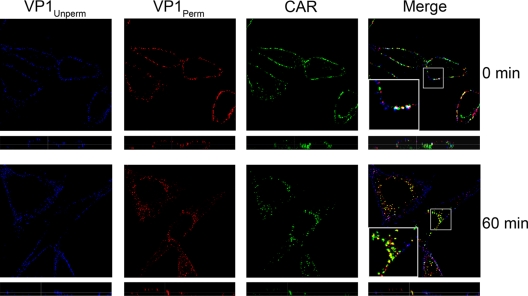
FIG. 2.
CVB3-RD enters with CAR. CVB3-RD particles were bound to cells and allowed to enter for 60 min as in Fig. 1C. Virus was detected with a blue fluorophore before permeabilization (VP1Unperm) and with a red fluorophore after permeabilization (VP1Perm). Uniquely red fluorescence indicates internalized virus. CAR was stained with a green fluorophore after permeabilization. Images were captured with a laser-scanning confocal microscope using an oil immersion 63× objective. Inserts within merge panels are magnified 3×.

