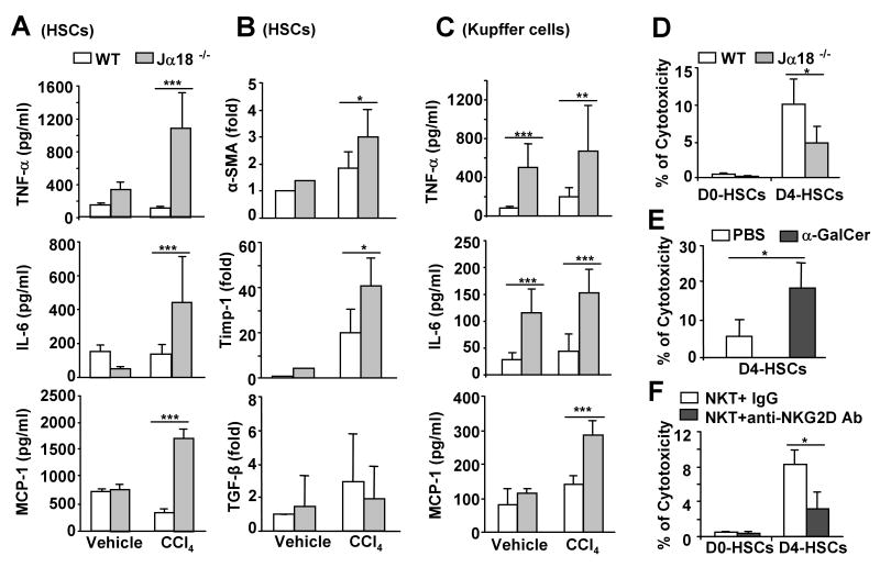
Fig. 3. Hepatic stellate cells (HSCs) from CCl4-treated Jα18-/- mice produce more cytokines and NKT cells kill early activated HSCs via an NKG2D-dependent mechanism.
HSCs and Kupffer cells were isolated from wild-type and Jα18-/- mice treated with CCl4 or vehicle (olive oil) for 12 h, and cultured in vitro for 12 h. A, The levels of cytokines from the supernatant of cultured HSCs. B. Expression of α-SMA, Timp-1, and TGF-β mRNA from the cultured HSCs. C. The levels of cytokines from the supernatant of cultured Kupffer cells. D. Liver lymphocytes from wild-type and Jα18-/- mice were incubated with freshly isolated HSCs (D0-HSCs) or 4-day cultured HSCs (D4-HSCs) for 4 h. HSC cell death was determined. E. Liver lymphocytes were isolated from mice treated with PBS or α-GalCer for 3 h. Cytotoxicity against D4-HSCs was determined. F. Purified liver NKT cells were incubated with D0- or D4-HSCs in the presence of IgG control or anti-NKG2D antibody for 4 h. The cell death of HSCs was determined. Values in panels represent means±SD from 3 independent experiments. *P<0.05, **P<0.01,***P<0.001.

