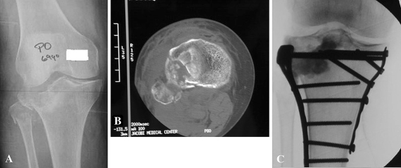
Fig. 1A–C.
(A) A plain radiograph of a tibial plateau fracture demonstrates the subchondral bone impaction. (B) An axial section of a computed tomography scan demonstrates the subchondral bone impaction in the tibial plateau fracture. (C) Intraoperative fluoroscopy demonstrates the anatomic reduction of the articular cartilage.

