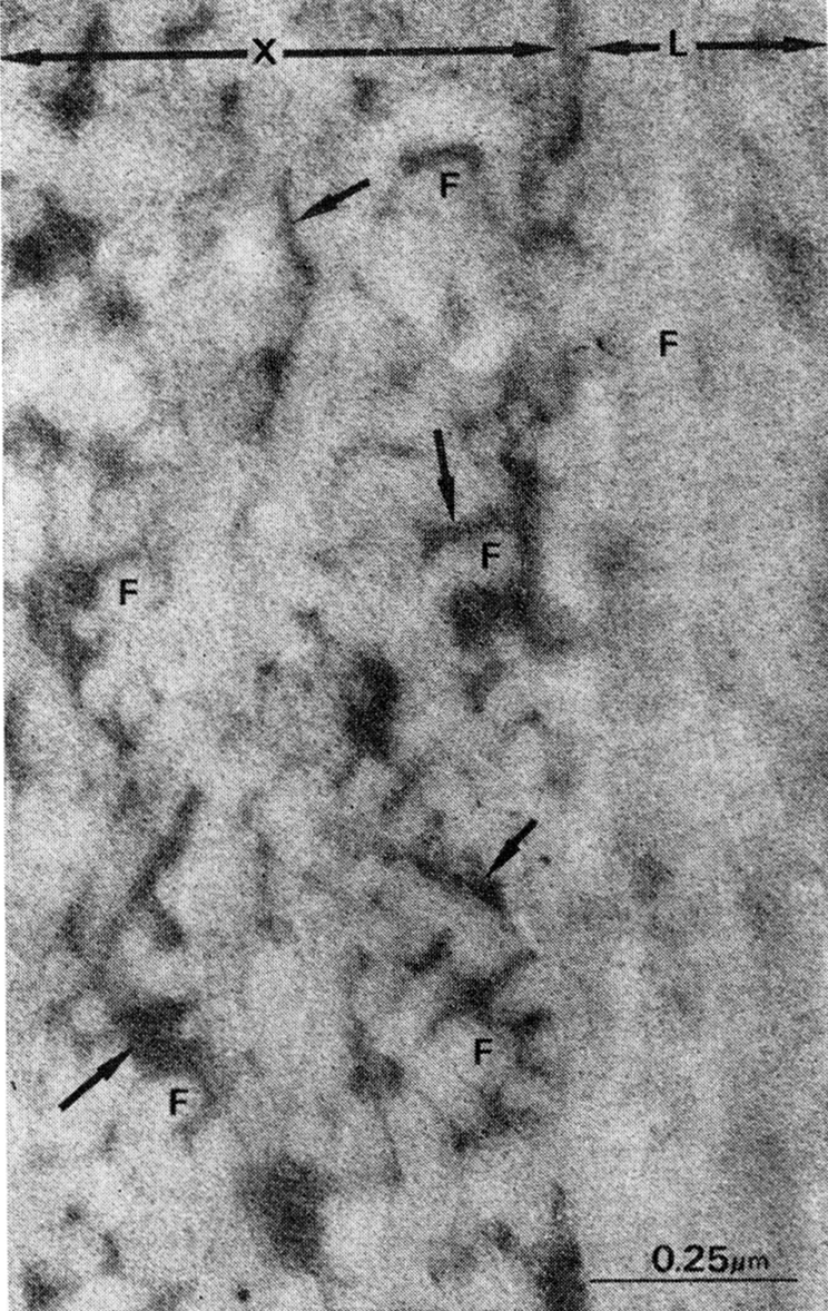
Fig. 1.

Demineralized cortical bone matrix, lyophilized and superfixed with 3% glutaraldehyde and 1.3% osmium tetroxide, cut in sections approximately 500 A thick and stained with uranyl acetate and lead citrate. Note large volume of space filled with interfibrillar electron-dense material, presumably proteoglycans (arrows) in the area cut in cross section (X). In longitudinal section (L) overlapping intertwined arrangement of collagen fibril (F) obscures large volume of interfibrillar noncollagenous material in bone matrix.
