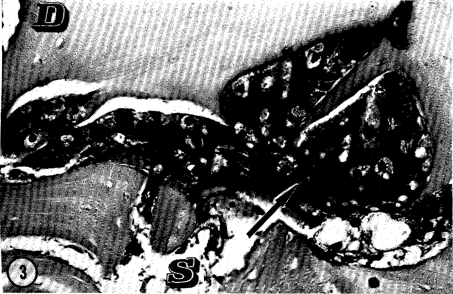
Fig. 3.
Photomicrograph of mature cartilage cells with abundant hyaline intercellular matrix, developed from explant of mesenchymal cell outgrowth of neonatal rat muscle onto the surface of decalcified bone matrix in BGJ media for 30 days. Old decalcified matrix (D), mesenchymal cells (S), and cartilage (arrow).

