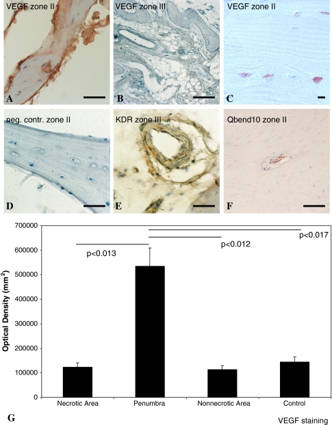
Fig. 4A–G.
Immunohistochemistry was performed to detect cells producing VEGF, VEGFR-2, or Qbend10. VEGF protein was immunostained in osteoblasts (A, C) from the penumbra and (B) from the periphery of necrotic femoral heads. (A, C) According to the staining intensity, VEGF was clearly upregulated in the case of steroid-related femoral head necrosis (ARCO Stage IV) in the penumbra of necrotic femoral heads. (D) Immunostaining is negative after preincubation of the primary antibody with recombinant human VEGF or after omission of the primary antibody (neg cont). (E) VEGFR-2 (KDR) could also be detected by immunohistochemistry in the periphery of necrotic femoral heads. (F) In the penumbra, Qbend10-positive immature vessels were detectable. (A, D, E, F) Bar = 20 μm; (C) bar = 100 μm (B); bar = 10 μm. (G) Densitometry of VEGF staining revealed strong upregulation of VEGF in the penumbra of ON femoral heads. Error bars = SD.

