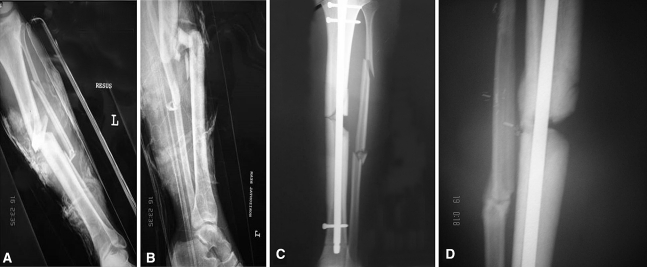
Fig. 1A–D.
Images illustrate the case of Patient 43 (Table 1) before combined treatment with autologous bone graft (ABG) and bone morphogenetic protein-7 (BMP-7). (A) Postinjury anteroposterior and (B) lateral radiographs show an open Grade IIIb tibial fracture. (C) A postsurgery anteroposterior radiograph shows the fracture after initial fixation (intramedullary nailing). (D) An anteroposterior radiograph taken 8 months postsurgery shows the evident atrophic nonunion.

