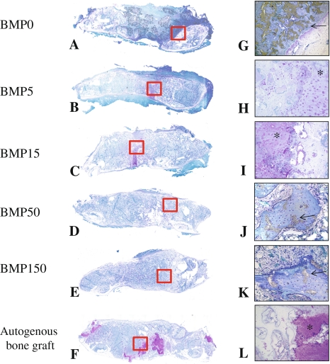
Fig. 4A–L.
Representative longitudinal histology sections of fusion mass of each group at 8 weeks after surgery are shown. (A) BMP0; (B) BMP5; (C) BMP15; (D) BMP50; (E) BMP150; (F) autogenous bone graft (Stain, Toluidine blue; original magnification, A–F: ×1). (G) BMP0; (H) BMP5; (I) BMP15; (J) BMP50; (K) BMP150; (L) autogenous bone graft (Metachromatic; original magnification ×100 for G and L, original magnification ×200 for H, I, J and K). Positive cartilage remnant in the fusion mass is not present in only section of groups of BMP50 and BMP150. More than 50 μg/side of E-BMP-2 treatment could histologically achieve lumbar spinal fusion as well as autogenous bone graft. Arrows indicate residual β-TCP.

