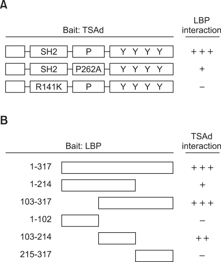
Figure 1.
Mapping by yeast two-hybrid assay of the LBP binding sites in TSAd (A) and the TSAd binding sites in LBP. Binding affinity was scored as +++ (deep blue), ++ (intermediate blue), + (weak blue) or - (white) upon X-Gal staining. SH2; Src homology domain, P; proline-rich motif, Y; tyrosine phosphorylation sites.

