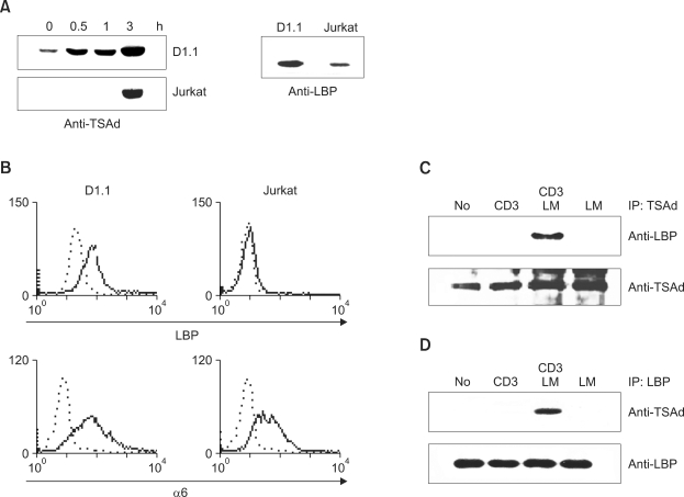
Figure 2.
TSAd associates with LBP in response to TCR + laminin (LM) stimulation. (A) D1.1 and Jurkat cells were stimulated with anti-CD3 for the indicated times, and cell lysates were subjected to Western blotting with anti-mTSAd Ab or anti-LBP Ab. (B) FACS histograms of D1.1 cells (left columns) and Jurkat cells (right columns) depicting surface expression of LBP and integrin α6. In each panel, staining with the indicated Ab is depicted by a solid line in comparison with an isotype control (broken line). (C) D1.1 cells were transfected with the expression plasmids encoding GFP-tagged TSAd and stimulated with anti-CD3 Ab, anti-CD3 Ab + laminin or laminin. Subsequently, GFP-tagged TSAd was immunoprecipitated with anti-TSAd Ab, and immunoprecipitates were subjected to western blotting with anti-LBP Ab and anti-TSAd Ab. (D) D1.1 cells were prepared as described in (C). Endogenous LBP was immunoprecipitated with anti-LBP Ab, and the immunoprecipitates were analyzed by Western blotting with anti-TSAd or anti-LBP Ab.

