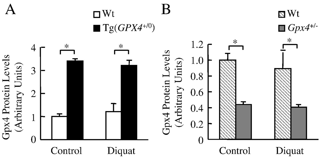
Figure 2. The levels of Gpx4 in Tg(GPX4+/0) and Gpx4+/− mice.
Tg(GPX4+/0) and Gpx4+/− mice and their corresponding Wt littermates were either untreated or treated with diquat. Gpx4 protein levels in the liver were determined by Western blotting as described in the Experimental Procedures. Panel A: Quantification of Gpx4 protein levels in the liver of Tg(GPX4+/0) mice (solid bars) and their Wt littermates (open bars) as determined from the Western blotting. Panel B: Quantification of Gpx4 protein levels the liver of Gpx4+/− mice (shaded bars) and their Wt littermates (hatched bars) as determined from the Western blotting. Values are mean ± SEM of data obtained from 3–4 mice. The asterisks indicate those differences that are significantly different.

