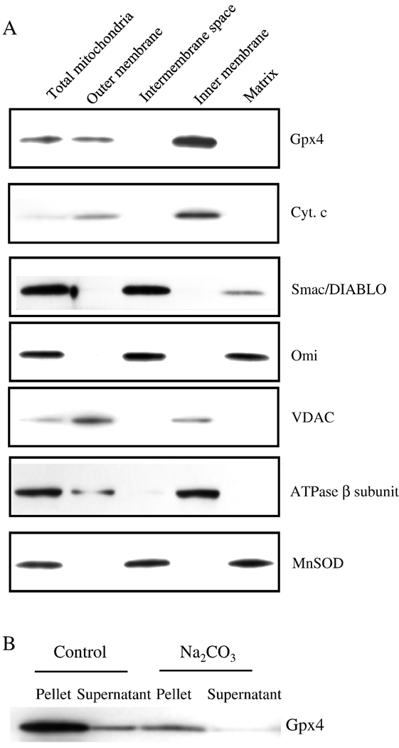
Figure 6. Submitochondrial localization of Gpx4 and apoptogenic proteins.
Submitochondrial fractions were obtained as described in the Experimental Procedures. Localizations of Gpx4 and apoptogenic proteins are shown by Western blotting. Panel A: Photograph of a representative Western blot showing the localization of Gpx4 and apoptogenic proteins in the mitochondria. Panel B: Photograph of a representative Western blotting showing the integral membrane association of Gpx4 with the inner membrane.

