
Fig. 3.
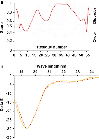
Circular dichroism evidence for natural unfolding of the murine extended N terminus. a Prediction of the disorder domain of the N-AChE terminus. b Shown is the profile of the 46 amino acids of the N-extended terminus of mouse AChE. Note the characteristic shape of an unfolded protein. Y axis: delta E (Delta e = e L − e R), showing the difference in extinction coefficients for left- and right-handed circularly polarized light. X axis: wavelength (yellows, predicted unfolded structure, red N-peptide)