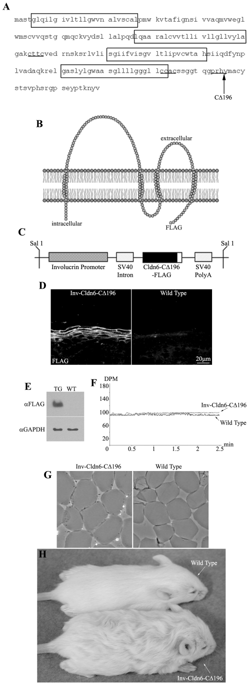
Figure 1. Inv-Cldn6-CΔ196 transgenic mice.
The Cldn6 protein sequence is shown; transmembrane-spanning regions are encased within boxes, the CXXC motifs are underlined, and the truncation site is indicated with an arrow (A). The Inv-Cldn6-CΔ196 mutant was created by deleting the C-terminal half of the cytoplasmic tail domain of Cldn6 after amino acid 196 (B). The Inv promoter was use to drive transgene expression to the suprabasal compartment of the transgenic mouse epidermis, where TJs are localized (C). Transgene expression was confirmed to be restricted to the upper spinous and granular layers of the epidermis as visualized by immunohistochemistry using anti-FLAG antibodies (D) and immunoblotting confirmed a ∼19.5kDa band corresponding to the transgene product in skin samples from transgenic (TG) but not wild type (WT) skin samples using anti-GAPDH as a loading control (E). Trans-epidermal water loss measurements confirmed no skin barrier dysfunction in the Inv-Cldn6-CΔ196 transgenic mice at birth (F). This was further supported by evaluation of cornified envelopes; which were characterized by a rigid shape and uniform size comparable to that of the wild type (G). Inv-Cldn6-CΔ196 mice were easily identifiable by their frizzed and lackluster coat appearance as compared to the wild type, a phenotype that persisted throughout life (H).

