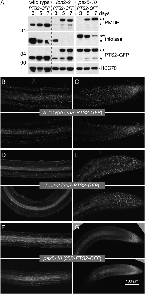
Figure 7.
lon2-2 roots have tissue-specific defects in PTS2-GFP import. A, Immunoblot of protein extracted from 3-, 5-, and 7-d-old wild-type (35S-PTS2-GFP), lon2-2 (35S-PTS2-GFP), and pex5-10 (35S-PTS2-GFP) seedlings (eight per lane). Seeds were stratified for 2 d prior to plating. Blots were probed with anti-PED1 (thiolase), PMDH2, GFP, and HSC70 antibodies. Positions of Mr markers (in kD) are indicated on the left; processed and unprocessed PTS2 proteins are marked by one or two asterisks, respectively. B to G, Root tip cells (C, E, and G) and maturing cells several millimeters above the tip (B, D, and F) of 6-d-old seedlings expressing PTS2-GFP were imaged via confocal microscopy. Micrographs are 11-μm-thick individual optical sections. Bar = 100 μm.

