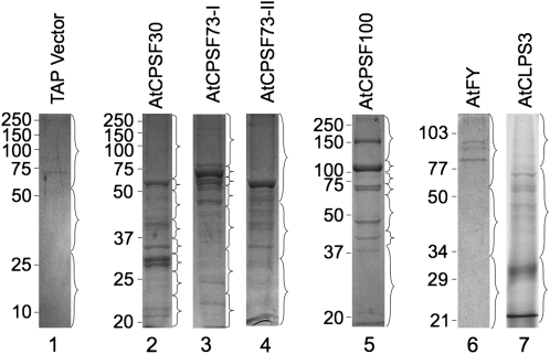
Figure 3.
Coomassie Brilliant Blue staining of proteins isolated by TAP purifications from Arabidopsis suspension cell cultures. The proteins were separated by 12% SDS-PAGE. Each gel lane was divided into a number of segments as indicated by the braces. The gel fragments were digested by trypsin overnight and subjected to LC-MS/MS for protein identification. Protein markers are as shown on the left (kD).

