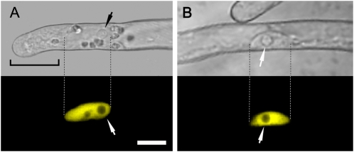
Figure 1.
Subcellular localization of the cameleon NupYC2.1 in M. truncatula wild-type root hairs. A, Elongating root hair with corresponding confocal fluorescence image showing that the NupYC2.1 labeling is specifically localized in the nucleus. Note that the nucleoli are devoid of fluorescence and that there is no detectable signal in the cytoplasmically dense tip region (bracket). B, Shank of a fully grown root hair in which the NupYC2.1-labeled nucleus is randomly positioned against the cell wall. Dashed lines indicate the nuclear position in corresponding bright-field and fluorescence images. The NupYC2.1 fluorescence is pseudocolored in yellow, and small arrows indicate the position of prominent nucleoli. The magnification is the same for all images. Bar in A is 15 μm.

