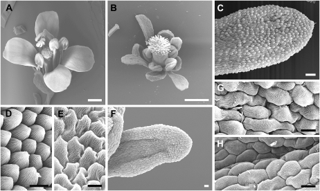
Figure 2.
Scanning electron micrographs showing defects in flower development in Pro35S:SPL/NZZ plants. A, A wild-type flower showing normal petals. B, A Pro35S:SPL/NZZ flower showing narrow petals and shortened stamens. C, A narrow petal in whorl 2 of a Pro35S:SPL/NZZ flower. D, Wild-type petal epidermal cells showing uniform size and conical shape. E, Wild-type anther epidermal cells exhibiting irregular cell edges and wavy ridges. F, A narrow and up-curled petal in whorl 2 of a Pro35S:SPL/NZZ flower. G, High-magnification view of epidermal cells of the organ shown in C displaying elongated shape and uneven sizes. H, High-magnification view of epidermal cells of the organ shown in F exhibiting the resemblance to wild-type anther epidermal cells in E. Bars = 0.5 mm (A and B), 50 μm (C and F), and 10 μm (D, E, G, and H).

