
Fig. 3.
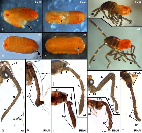
Parental RNAi with Sp8/9 in Oncopeltus. a, b Embryos after Sp8/9 RNAi shortly before hatching in lateral (a) and ventral (b) aspect. c Hatchling after Sp8/9 RNAi. d, e Wild-type embryos shortly before hatching in lateral (d) and ventral (e) aspect. f Wild-type hatchling. The arrows in (a) and (d) denote the tips of the legs. The arrows in (b) and (e) denote the tips of the antennae. All animals in (a)–(f) are oriented with the anterior end to the left. g–m Preparations of the appendages of wild-type hatchlings (g, i, k) and hatchlings after Sp8/9 RNAi (h, j, l, m). The wild-type rostrum (g) comprises the four-segmented labium, filiform maxillae and mandibles, and the pointed labrum. The rostrum of RNAi animals (h) is malformed with fused distal segments. The wild-type antenna (i) comprises an antennifer and four additional segments. The antenna of RNAi animals is severely shortened with fused segments (j). The arrow in (j) points to a small ectopic outgrowth on the antenna seen in about half of the hatchlings after Sp8/9 RNAi. The wild-type legs (k) comprise a coxa, trochanter, femur, tibia, and a two-segmented tarsus with two claws. The legs of RNAi animals are shortened with fused tarsal segments (l, m). The more proximal segments coxa, trochanter, and femur may be partially (l) or fully fused together (m). Abbreviations: lr labrum, an antenna, af antennifer, ro rostrum, md mandible, mx maxilla, lb labium, t1 prothoracic leg, t2 mesothoracic leg, t3 metathoracic leg, cx coxa, tr trochanter, fe femur, ti tibia, ta tarsus, cl claw