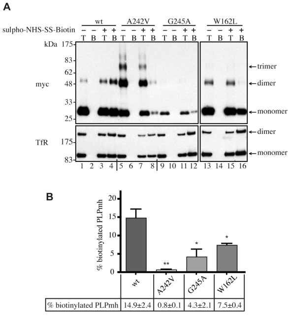
Fig. 3.
Analysis of cell-surface expression of wt and mutant PLP. (A) HeLa cells expressing wt or mutant PLPmh were left untreated (–) or were treated (+) with membrane-impermeant sulpho-NHS-SS-biotin. Biotinylated proteins were precipitated using NeutrAvidin beads (beads, `B') and analysed by immunoblotting with anti-Myc or anti-TfR antibodies. The biotinylation levels of wt and mutant PLPmh were compared with 20% total cell lysates (total, `T'). (B) The levels of monomeric and oligomeric PLPmh on immunoblots were quantified by densitometry. Biotinylated PLPmh was expressed as a percentage of total PLPmh and normalised to the equivalent TfR levels. Histogram shows mean ± s.e.m. from three independent experiments (*P<0.05, **P<0.01, t-test).

