Fig. 2.
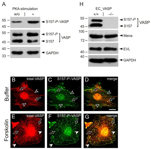
VASP translocation to the cell periphery depends on S157 phosphorylation. Wild-type endothelial cells (EC_VASP+/+) were incubated with forskolin (5 μM) or buffer and analyzed using antibodies against S157-P–VASP and total-VASP. (A) Western blots of cell lysates normalized for GAPDH. (B-G) Immunofluorescence images of fixed and permeabilized cells. The merge of total-VASP (red; B,E) and S157-P–VASP (green; C,F) is given in yellow (D,G). Black arrowheads indicate stress fibers, black arrows indicate focal adhesions, and white arrowheads indicate plasma membranes. Images were taken with a 100× objective and are representative of a series of eight experiments. Scale bar: 15 μm. (H) Comparison of VASP, Mena and EVL expression in EC_VASP–/– and EC_VASP+/+ cells by western blotting. The cell lysates are normalized for GAPDH.

