
Figure 5. X-band electron paramagnetic resonance (EPR) spectra showing displacement of CN- by NO at heme a3 of cytochrome c oxidase.
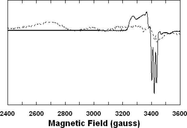
Sample preparations were carried out in 0.1 M aqueous potassium phosphate buffer, pH 7.4, 1.0 mM in EDTA, 0.05% in lauryl maltoside, 22 °C, prior to freezing in EPR tubes. Recording conditions: 0.2 mW microwave power, 4 G modulation amplitude, 1 × 10⁴ amplifier gain, 15 K sample temperature. Broken trace: partially-reduced cyanide adduct, 60 μM in enzyme, 1.0 mM in KCN, ~1 mM in Na2S2O4. Solid trace: partially-reduced cyanide adduct plus NO, 60 μM in enzyme, 1.0 mM in KCN, ~1 mM in Na2S2O4, 1.9 mM (1.0 atm) NO.