
Figure 1.
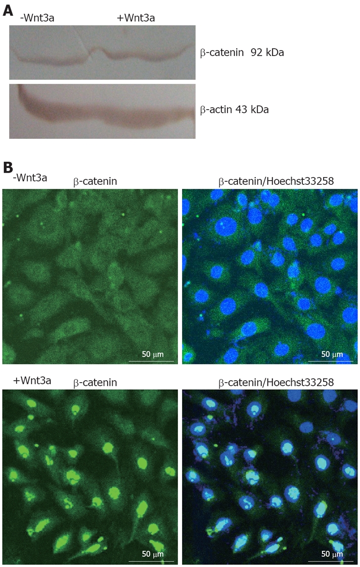
Effects of Wnt3a on β-catenin expression, its subcellular localization, and induction of typical Wnt target genes. A: Stimulation of WB-F344 cells with 160 ng/mL Wnt3a for 1 d revealing a slight increase in β-catenin protein level as shown by Western blot analysis and densitometric analysis; B: Immunocytochemistry analysis of β-catenin exhibiting perinuclear staining for β-catenin in unstimulated WB-F344 cells (upper panels), whereas addition of 160 ng/mL Wnt3a for 1 d (lower panels) showing clear nuclear staining for β-catenin. Immunofluorescence was performed using a polyclonal antibody against β-catenin (left panels). In addition, nuclei of WB-F344 cells were stained with Hoechst33258 (right panels). Scale bars: 50 μm.