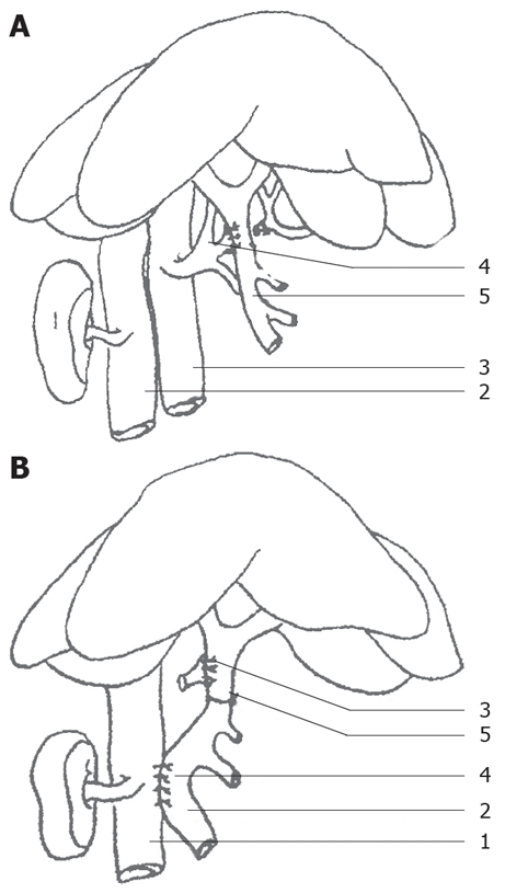
Figure 1.

Animal PVA model establishment. A: Side-to-side anastomotic stoma of the hepatic artery and the portal vein. (1) kidney; (2) inferior vena cava; (3) abdominal aorta; (4) hepatic artery; (5) portal vein. B: Side-to-side anastomotic stoma of the portal vein and the venae cavae. (1) inferior vena cava; (2) portal vein; (3) arteriovenous anastomotic stoma; (4) anastomotic stoma of the portal vein and the venae cavae; (5) ligation of the portal vein.
