
Figure 5.
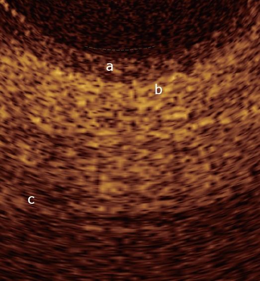
Magnification of an OCT image from the normal main pancreatic duct wall. From the surface of the duct, up to a depth of 1 mm, the following layers are recognizable: The single layer of epithelial cells, approximately 0.04-0.08 mm thick, visible as a superficial, hypo-reflective band (a); the connective-fibro-muscular layer surrounding the epithelium, visible as a hyper-reflective layer approximately 0.36-0.56 mm thick (b); the connective and acinar structure close to the ductal wall epithelium, visible as a hypo-reflective layer (c).