
Figure 9.
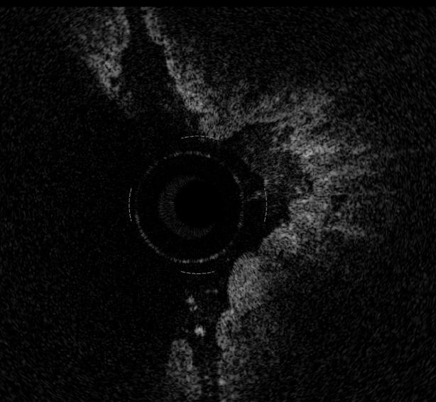
Magnified OCT imaging of early stage adenocarcinoma raised within Barrett's epithelium. The lack of the regular esophageal wall layered morphology and a markedly heterogeneous back-reflectance of the signal characterize the neoplastic lesion, which is confined within the epithelium.