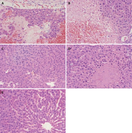
Figure 1.
Pathological changes of liver under light microscope. A: Lamellar hemorrhagic necrosis, model control group (6 h); B: Massive hemorrhagic necrosis, model control group (12 h); C: Spotty necrosis of liver cell, Baicalin treated group (12 h); D: Piecemeal necrosis, Octreotide treated group (12 h); E: Normal liver, sham-operated group (12 h). (HE, × 200).

