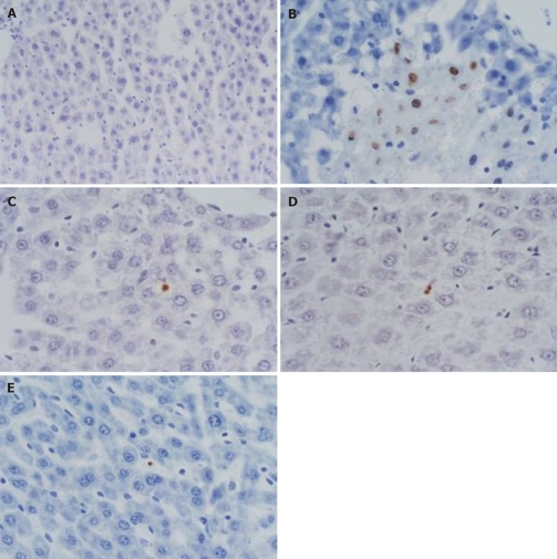
Figure 3.
TMA of liver was prepared and conduced TUNEL staining, observing the changes of apoptotic indexes. A: model control group (3 h), there was no apoptotic cell; B: Baicalin treated group (3 h), several hepatic apoptotic cells appeared; C: Baicalin treated group (6 h), several apoptotic hepatic Kupffer cells; D: Baicalin treated group (12 h), several apoptotic hepatic Kupffer's cells; E: Octreotide treated group (3 h), several apoptotic hepatic Kupffer's cells. (TUNEL, × 400).

