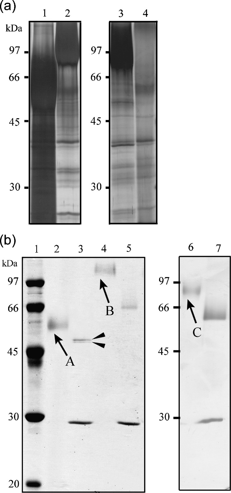
Fig. 3.
SDS-PAGE profile using culture media (a) and purified recombinant proteins from transformants as samples (b). The proteins contained in culture media and the purified proteins were separated using a 10% SDS gel and visualized with silver nitrate or Coomassie Brilliant Blue. a Media from the culture of A89CS3ocpA (lane 1), A89CS3ocpB (lane 2), A89CS3cpI (lane 3), and A. nidulans A89 (lane 4). bArrowed bands were excised from the gel for identification by peptide mass fingerprinting. Bands appeared at 30 kDa in lanes 3, 5, and 7 correspond to endoglycosidase H. Lane 1 Molecular weight marker; lanes 2, 4, and 6 purified recombinants OcpA, OcpB, and CpI, respectively; lanes 3, 5, and 7 deglycosylated purified recombinants OcpA, OcpB, and CpI, respectively. Deglycosylated recombinant OcpA was shown as two bands (filled arrowheads)

