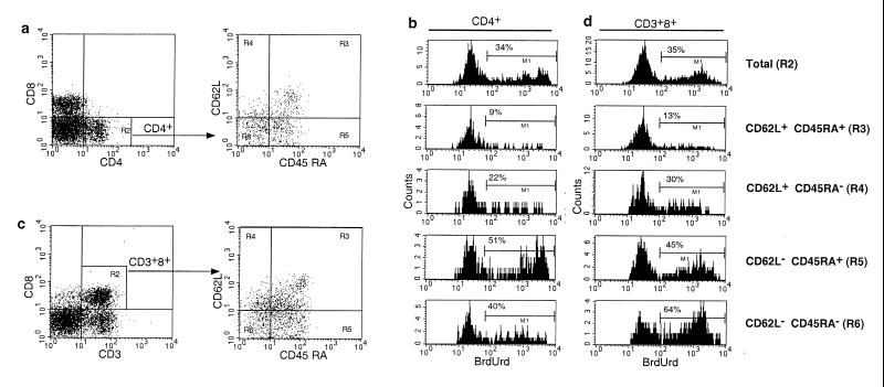
Figure 1.
Five-color flow cytometry analysis of in vivo BrdUrd labeling in an SIV-infected macaque. Peripheral blood lymphocytes from an SIV-infected rhesus macaque that had been labeled with BrdUrd were surface stained with antibodies to CD3, CD8, CD45RA, and CD62L or CD4, CD8, CD45RA, and CD62L, then permeabilized, and stained with antibodies to BrdUrd. (a) Gating of CD4+ T lymphocytes. Cells enclosed in the gate designated by R2 then were analyzed for expression of CD45RA and CD62L. (b) Analysis of BrdUrd uptake in memory and naive populations of CD4+ T cells. (c) Gating of CD8+ T lymphocytes. To exclude CD3–CD8+ natural killer cells, subsequent analysis of expression of CD45RA, CD62L, and BrdUrd was confined to CD3+CD8+ cells indicated in the R2 gate. (d) Analysis of BrdUrd uptake in memory and naive populations of CD8+ T cells. The gate corresponding to each population of naive and memory cells is indicated in parentheses. The percentage of BrdUrd-labeled cells for each gated cell population is shown in each histogram.

