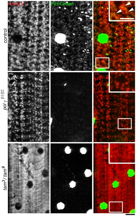
Figure 1. Mutations in the catalytic and accessory subunits of DNA polymerase γ impair mtDNA replication and decrease mtDNA content.
An antibody against mitochondrial complex V (red) and the dye PicoGreen (green) are used to label mitochondria and mtDNA respectively, in muscles of wildtype control, pol γ β1/β2 and tam 3/tam 9 crawling 3rd instar Drosophila larvae. Regular mitochondrial distribution is disrupted in pol γ β1/β2 and tam 3/tam 9 mutants and number of mtDNA nucleoids are significantly reduced. PicoGreen also labels the dsDNA of muscle nuclei that serves as internal control for the staining. Muscle nuclei appear smaller in the pol γ mutants. Because of the relatively high concentration of dsDNA in muscle nuclei, they appear saturated at offset levels required to visualize the smaller mtDNA nucleoids. Insets in the RGB merge show digitally magnified regions from the boxes and arrowheads indicate presence of mtDNA in control muscles and are absent in muscles of pol γ mutants. tam 3/tam 9 larvae have visibly higher mitochondrial density. Scale bars equal 10 µm.

