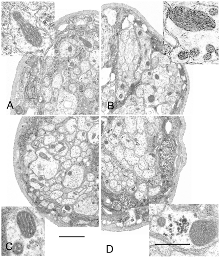
Figure 6. Mitochondrial ultrastructure in segmental nerves.
Each quadrant shows a portion of a segmental nerve from each of the four types of animals that were examined. No differences in the nerve structure were noticed between control and mutant larvae. (A) Background control (UAS-mtGFP; D42-Gal4 in wildtype background); (B) Canton S; (C) tam 3/tam 9; and (D) pol γ-β1/β2. Cross-sections of mitochondria (insets) are found in about 1 out of every 5 axonal profiles in controls. The relative number of mitochondrial profiles in axons appeared to be slightly increased in pol γ-β1/β2 and tam 3/tam 9 mutants, to around 1 out of 3 in a sample of 3 nerves each. Oblique or longitudinal sections show that axonal mitochondria can be quite long and threadlike, and range in diameter from 100 to 800 nm. Both large and small diameter mitochondria may be found side by side in the same axon (B, C, D). Glycogen granules are seen in some of the large diameter axons (D). Scale bars equal 1 µm for nerve quadrants and 0.5 µm for insets.

