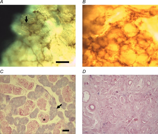
Figure 1.
Control and ligated submandibular glands Nerve staining of collagenase-digested cell clumps from control (A) and 4-week-ligated glands (B). Cholinesterase nerve staining indicates that parasympathetic nerves (arrow in A) are still attached following collagenase digestion. Alcian Blue/Periodic Acid Schiff's staining of tissue sections reveals blue acinar cells (arrows) and pink granular ducts (*) in normal glands (C) and an almost complete loss of secretory granules in 4-week-ligated glands (D). Scale bar represents 30 μm.

