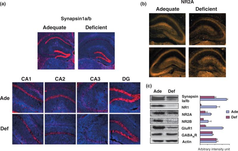
Fig. 6.
Localized reduction of synapsins and NR2A in young mouse hippocampi depleted with DHA during development. (a) Immunohistochemical probing of synapsin1 and NR2A in P-18 hippocampal slices, showing dramatic decreases of synapsin1 expression in CA1, CA2, and CA3 regions from DHA-depleted (Def) mice in comparison to DHA-adequate (Ade) mice; synapsin1 in red and DAPI counterstaining in blue. (b) Immunohistochemical probing of NR2A in P-18 hippo-campal slices from DHA-adequate (Ade) or DHA-depleted (Def) mice, indicating particular decrease of NR2A in the CA3 region of DHA-depleted hippocampi. (c) Western blot analysis of synapsin1, NR, GluR and GABA receptor expression in hippocampi of DHA-adequate or deficient mice at P-18.

