
Figure 7. Localization of DNA and SeqA in cells irradiated with UV.
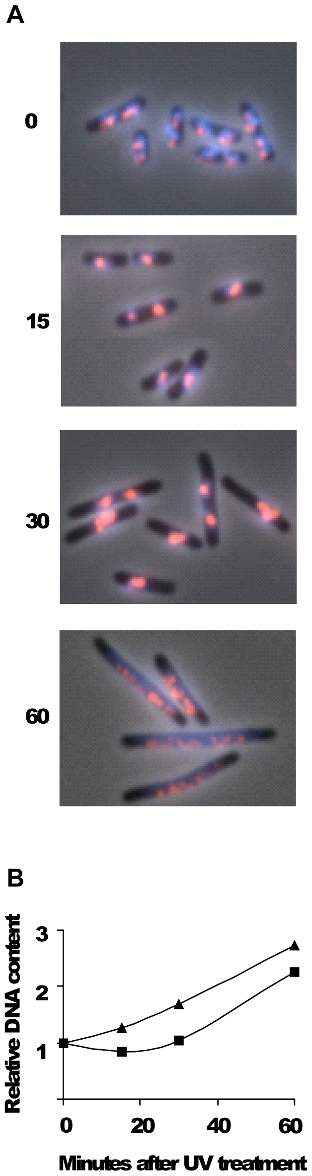
MG1655 wild type cells were grown exponentially in glu-CAA-uri medium at 37°C to OD450 = 0.15 and then irradiated with UV (50 J/m2). Samples were taken at the time points indicated and fixed in 70% ethanol. A: Cells were immunostained with antibody against SeqA (red) and the DNA was stained with Hoechst 33258 (blue). The two fluorescence images are shown merged with the phase-contrast image. B: Relative DNA content per cell obtained from flow cytometry analysis as a function of time after UV treatment. One portion of exponentially growing cells (OD450 = 0.15) was exposed to UV while another portion was mock-treated, then 10 µg/ml cephalexin was added to both cultures and samples taken at the time indicated. ▴: mock-treated cells, ▪: irradiated cells. The data in panel B are the average of two independent experiments. Samples in panel A were not treated with cephalexin.