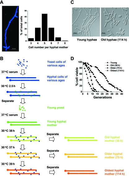
Fig. 2.
Large-scale separation of old and young cells. (A) Most of the hyphae induced under the experimental conditions contain four to six single hyphal cells. Left panel, cells stained by Calcofluor and photographed under microscope; right panel, statistical results. (B) Schematic procedures for large-scale preparation of old cells. (C) Morphological differences between old and young cells. Photos were taken with a Zeiss Axioplan 2 microscope using 10 × 100 magnification. (D) Lifespan analysis of hyphal cells separated at the time points of 0 h, 38 h, 75 h and 114 h, respectively. Mean lifespan and sample size are: 22.9 (n = 76), 13.1 (n = 76), 8.4 (n = 74) and 5.4 (n = 56), respectively.

