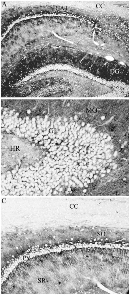
Fig. 2.
Sagittal section of rat brain hippocampus immunolabeled for NHERF1. (A) Strong labeling was observed in grey matter regions, including dentate gyrus (DG) and CA1 region (CA1), while negligible labeling was observed in white matter regions such as corpus callosum (CC). (B) In dentate gyrus, strongest labeling was observed in the molecular layer (MO), whereas weak labeling was observed in the hilar region (HR). In granule cell layer (GL), significant labeling was associated with astrocytes surrounding unlabeled neuronal elements. (C) In CA1 region, the strata oriens (SO) was strongly labeled, while less staining was observed for the strata radiatum (SR). Significant labeling was associated with astrocytes surrounding unlabeled neuronal elements such as pyramidal cells (py). Scale bars: (A) 100 μm, (B, C) 25 μm.

