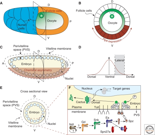
Figure 1.
Overview of the ventral signaling pathway. (A) Schematic of St. 10 egg chamber. The oocyte nucleus is located at the dorsoanterior cortex. Gurken, which is locally translated, is present in a protein gradient (green). (B) Cross section of schematic of St. 10 egg chamber. Gurken signaling (green) represses pipe expression (brown) in the follicle cells. (C) Schematic of syncytial blastoderm embryo. The ventral follicle cells, which had expressed pipe, deposit an unknown “chemical asymmetry” into the perivitelline space. (D) The chemical asymmetry results in a ventral-to-dorsal signaling gradient. (E) Cross section of schematic of syncytial blastoderm embryo. The signaling gradient is initially established within the perivitelline space, a small extracellular space between the embryo and an outer vitelline membrane. (F) Illustration of ventral signaling pathway in the early embryo. In the perivitelline space (PVS), a protease cascade (Ndl, Gd, Snk, Ea) eventually activates Spz, the ligand for the Toll receptor. The serine protease inhibitor, Spn27a, inhibits the activity of Ea. Activated Spz transduces the signal into the embryo through Toll, causing the degradation of Cactus and the nuclear translocation of the transcription factor Dorsal. The roles of Tube (T), Pelle (P), Weckle (W), and Myd88 (M) are relatively unknown, but participate in a signaling complex at the cytoplasmic tail of Toll.

