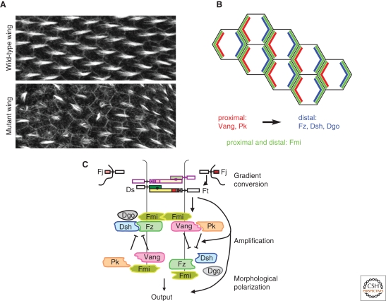
Figure 1.
Planar cell polarity in Drosophila. (A) Image of wild type (top panel) and PCP mutant Drosophila pupal wing epithelium, labeled with phalloidin to stain actin. (B) Schematic of PCP protein asymmetric cortical distribution in the fly wing epithelium showing Pk and Vang enriched on the proximal, Fz, Dsh, and Dgo on the distal and Fmi on both proximal and distal sides of each cell. (C) A model for organization of the PCP pathway in Drosophila. Heterodimers of Ft and Ds show biased orientation at each cell boundary, resulting from graded expression of Fj and Ds. Asymmetrically oriented Ft-Ds heterodimers bias the function of a feedback loop consisting of the core PCP proteins, Fmi, Fz, Dsh, Dgo, Vang, and Pk.

