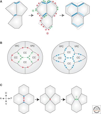
Figure 4.
Remodeling cell–cell junctions during morphogenesis. (A) Hexagonal packing in fly wing epithelial cells. Remodeling involves cell–cell contact shrinking (red arrows), expansion (green arrows), loss of some contacts (red asterisk), and creation of new contacts (green asterisk). The previous shape of cells is indicated by dashed lines in the middle and right panels. Flamingo enrichment (dark blue rectangles) and emergent polarity (light blue rectangles) may spatially bias exocytic delivery of E-cad to promote hexagonal packing. (B) Pattern formation in fly retina. (Left panel) Wild type ommatidial cluster with four cone cells (CC) surrounded by two primary pigment cells (1PC). Strong adhesion (N-cad + E-cad) increases inter-CC contacts (green arrows) at the expense of weaker adhesion (E-cad only) with 1PCs (red arrows). (Right panel) Uniform adhesion in a mutant ommatidium (blue arrows) distorts the cell pattern. (C) Cell intercalation during germ band elongation in fly embryos. The T1 transition involves shrinking of junctions between A/P neighbors (vertical junctions) and creation of new junctions between D/V neighbors (horizontal junctions). Shrinking is triggered by increased acto-myosin tension along A/P junctions (red arrows; vertices are indicated by blue dots). Expansion of the new D/V interface could rely on adhesion (green arrows), as in cell culture systems.

