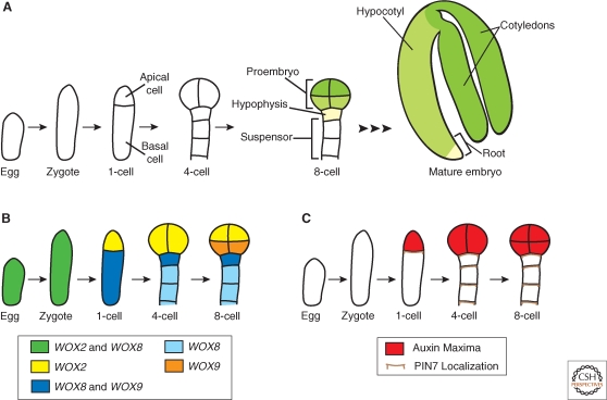
Figure 2.
Embryo development and the asymmetric localization of factors in the embryo. (A) Schematic of embryo development focusing on the events from the egg to the eight-cell stage. After fertilization, the zygote expands and then divides asymmetrically to produce the apical and basal cells. Embryo stages are based on the number of cells in the apical domain only, thus the first zygotic division results in a one-cell stage embryo. The apical cell undergoes a series of divisions to generate the eight-celled proembryo. The basal cell undergoes a series of strictly transverse divisions to generate the suspensor. Later in development, the uppermost cell of the suspensor, the hypophysis, is incorporated into the embryo. Therefore, by the eight-cell stage, four anatomically distinct cell types are present: The upper (green) and lower (yellow-green) tiers of the proembryo, the hypophysis (cream), and the suspensor (white). The suspensor remains extraembryonic, whereas the remaining cell types give rise to the cotyledons, hypocotyl, and root of the mature embryo as depicted by the corresponding color scheme in the eight-cell stage and mature embryo. (B) Schematic of the WOX gene expression patterns. By the eight-cell stage, the expression domains of the WOX genes coincide with the four distinct cell types present. (C) Schematic of the auxin maxima and PIN7 localization. PIN7 localization on the upper membrane of basal cells directs the flow of auxin from basal to apical cells. This polarized movement of auxin generates an auxin maximum in the apical domain as determined by expression of the DR5 reporter.

