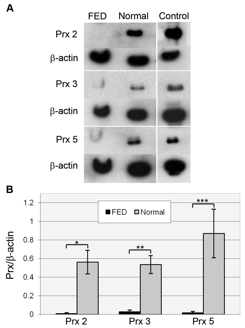
Figure 2.
Western blot analysis of peroxiredoxin (Prx) isoform expression in normal and FED HCEC-DM complexes. (A) Representative Western blots compare expression of Prx-2, -3, and -5 in FED and normal corneal endothelial samples. Positive controls included LNCap cell lysate for Prx-2 and HeLa cell lysate for Prx-3 and -5. Beta-actin was used for normalization of protein load. (B) Densitometric comparison of the average expression of Prx-2, -3, and -5. Bars: SEM. *: p=0.0045; **: p=0.0080; and ***: p=0.011.

