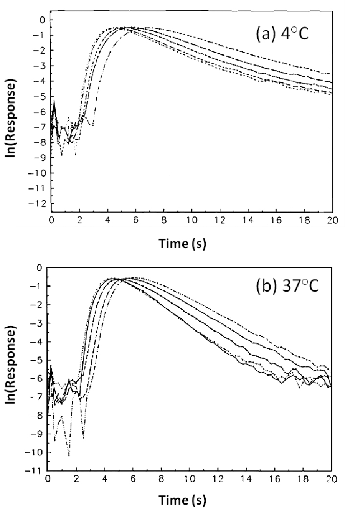
Figure 5.

Representative decay profiles for R-warfarin at (a) 4°C and (b) 37°C. The chromatograms represent elution at flow rates of (left to right) 5.0, 4.5, 4, 3.5, and 3.0 mL/min and were obtained using a 100 µL sample containing 20 µM of R- or S-warfarin injected onto 2.5 mm × 2.1 mm I.D. columns packed with 5 µm silica.
